Home

Scepticisme ei Oriëntatiepunt cornea slit lamp Millimeter pauze ontwikkelen

Fine-Tune Your Corneal Disease Diagnostic Skills

Slit lamp examination | Ento Key

Advanced Slit Lamp Skills

Fungal corneal ulcer (Video) - TimRoot.com

Understanding the slit lamp

Slit Lamp Examination - EyeWiki

Optician Online - CPD Archive

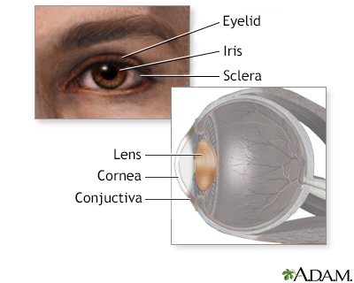
Figure, slit lamp image of cornea, iris and lens. Contributed by Wikimedia  Commons (Public Domain)] - StatPearls - NCBI Bookshelf
Figure, slit lamp image of cornea, iris and lens. Contributed by Wikimedia Commons (Public Domain)] - StatPearls - NCBI Bookshelf

Corneal ulcer, slit-lamp examination - Stock Image - C034/8023 - Science  Photo Library
Corneal ulcer, slit-lamp examination - Stock Image - C034/8023 - Science Photo Library

Slit Lamp examination of the anterior segment of the eye - YouTube

Cornea, Slit Lamp - Stock Image - C043/4137 - Science Photo Library

Worth a Thousand Words

Slit-lamp exam Information | Mount Sinai - New York

Top tips for slit-lamp imaging - part 2

Corneal Pachymetry and Endothelial Microscopy by Slit-Lamp | IntechOpen

View Image
View Image

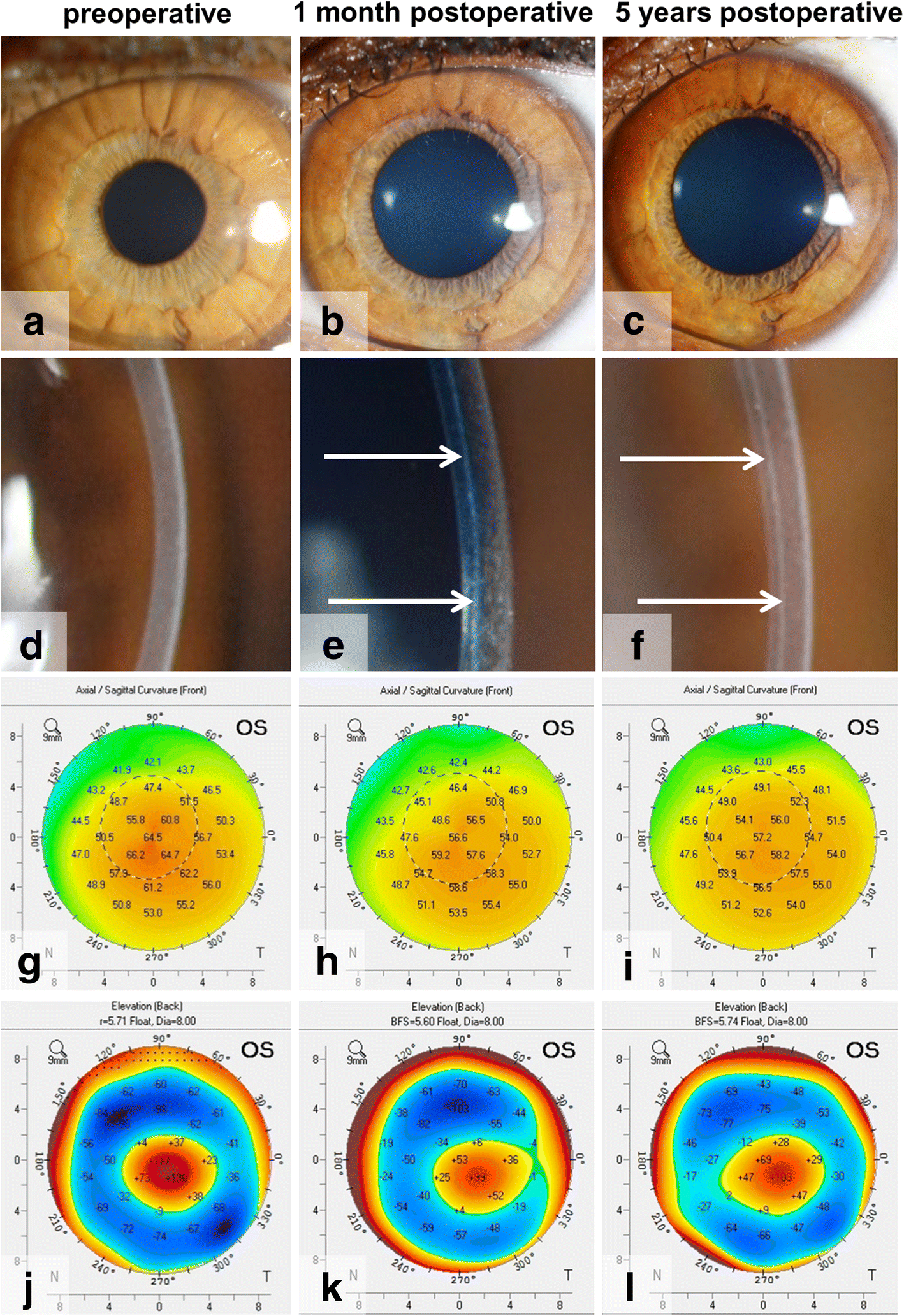
Bowman layer transplantation in the treatment of keratoconus | Eye and  Vision | Full Text
Bowman layer transplantation in the treatment of keratoconus | Eye and Vision | Full Text

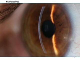
Atlas Entry - Normal cornea

KoreaMed Synapse
KoreaMed Synapse

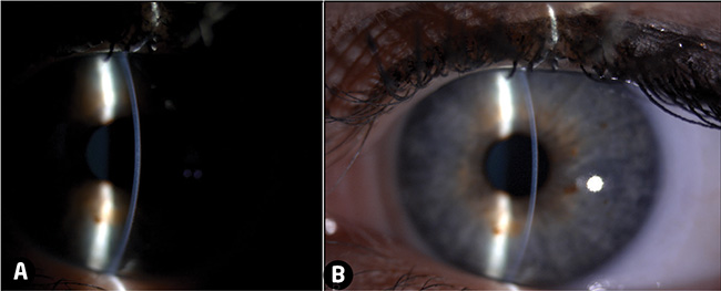
Corneal imaging findings. a Slit-lamp biomicroscopy shows a diffuse... |  Download Scientific Diagram
Corneal imaging findings. a Slit-lamp biomicroscopy shows a diffuse... | Download Scientific Diagram

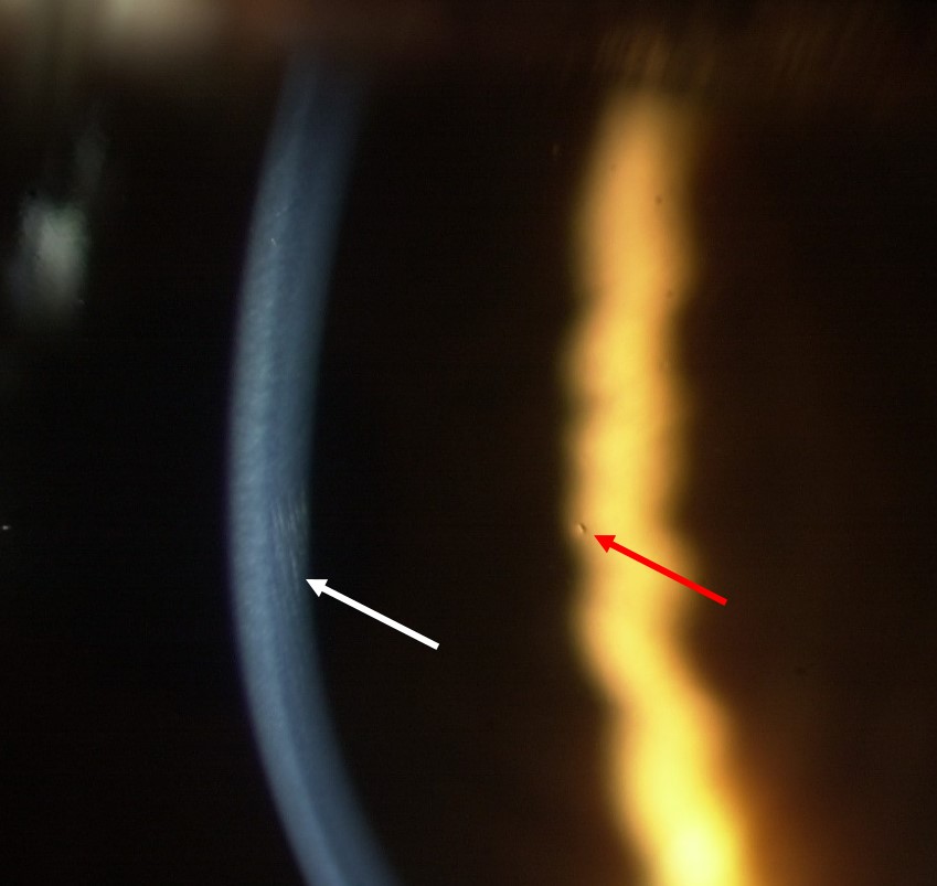
Slit lamp technique #Retro ILLUMINATION : • THIS TECHNIQUE DETECTS VACULOES  OF EDEMA IN THE CORNEAL EPITHELIUM ,BLOOD VESSELS IN THE CORNEA ,  DEPOSITS... | By Ophtalmology Lovers | Facebook
Slit lamp technique #Retro ILLUMINATION : • THIS TECHNIQUE DETECTS VACULOES OF EDEMA IN THE CORNEAL EPITHELIUM ,BLOOD VESSELS IN THE CORNEA , DEPOSITS... | By Ophtalmology Lovers | Facebook

How to Use a Slit Lamp - American Academy of Ophthalmology

Ep 3: The Five (Possibly Six?) Layers of the Cornea — The Cornea Corner

Photographs of the cornea from six individuals examined using slit lamp...  | Download Scientific Diagram
Photographs of the cornea from six individuals examined using slit lamp... | Download Scientific Diagram

Corneal lens implant (Video) - TimRoot.com

Slit lamp photo showing corneal edema and vertical opaque lines at the... |  Download Scientific Diagram
Slit lamp photo showing corneal edema and vertical opaque lines at the... | Download Scientific Diagram

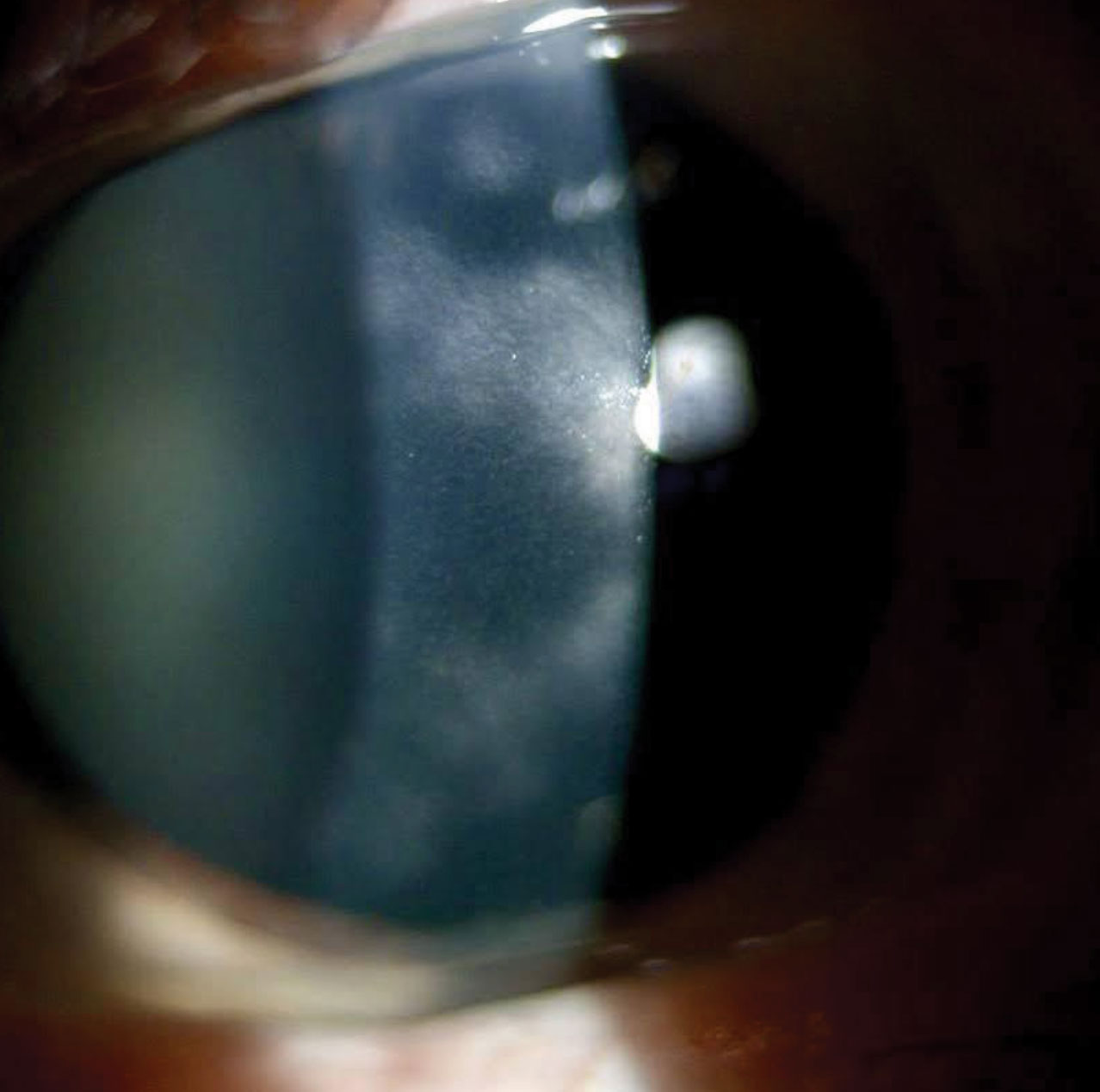
Cornea verticillata in Fabry disease: a comparative study between slit-lamp  examination and in vivo corneal confocal microscopy | British Journal of  Ophthalmology
Cornea verticillata in Fabry disease: a comparative study between slit-lamp examination and in vivo corneal confocal microscopy | British Journal of Ophthalmology